Professional Gynecological Services
- *Same day appointments & walk-ins welcome
- (718) 875-4848
- Book Online
Professional Gynecological Services
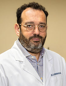
Dr. Dmitriy Bronfman, MD, is a board-certified obstetrician-gynecologist with over 25 years of experience providing complete gynecological care. He is an expert in many aspects of modern women's health, including preventative medicine, pelvic pain, minimally invasive and robotic surgery, and general, adolescent, and menopausal gynecology.
Dr. Bronfman graduated magna cum laude from New York University and received his medical degree from Mt. Sinai School of Medicine before completing his residency at Brooklyn Hospital Medical Center. He currently practices at Professional Gynecological Services and is affiliated with Lutheran Medical Center, New York Methodist Hospital, and The Brooklyn Hospital Center. Dr. Bronfman, together with the core physicians of Professional Gynecological Services, implements state-of-the-art diagnostic equipment, the most tested treatments, and the latest surgical technology, all while establishing a welcoming atmosphere in which your questions and concerns will be addressed with the utmost attention.
More about our teamThe Women’s Choice is an ACR Accredited Facility for ultrasound. We provide the highest quality imaging and patient safety, ensuring you receive world-class diagnostic care.